Cells, genes and the making of adult form
How genomes build adult organisms and how variation in mechanism produces difference in form and function — questions we ask using pigmentation and other traits
UNIVERSITY OF VIRGINIA | Department of Biology | Charlottesville, Virginia


parichylab development and evolution pigment pattern zebrafish regeneration
Our research
We study how adult phenotypes are built by cells and genes, and how variation in underlying mechanisms produces differences between individuals and species. The same processes that generate normal variation, when perturbed, give rise to birth defects and disease.
We are interested in how cells acquire and change identity, how they signal and migrate, how stem cells are maintained and recruited, how endocrine, paracrine and environmental cues coordinate growth and differentiation across tissues, and how all of this is rewired over evolutionary time to produce phenotypic novelty.
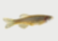
As a model, we often use pigment cells and pattern formation in zebrafish and other fishes; this system is biomedically important and unusually tractable, with chromatophores visible at single-cell resolution in living animals and patterns that vary spectacularly across species. But we also study other traits where the same questions can be asked productively during normal development, in regeneration, and when either goes awry: skin and scale development, integrated programs that build adult tissues, and lineages that maintain and regenerate them.
Our approaches include forward genetics and CRISPR mutagenesis and editing, single-cell transcriptomics, super-resolution time-lapse imaging, comparative genomics, biochemistry, and behavior. This breadth follows from the questions, since in the systems we study cells, genes, environment, and evolution are really not separable, and the most interesting problems tend to sit where they meet.
Three themes, one program


1 |
Pattern
formation
How do cells organize themselves into reproducible spatial arrangements? Stripes, spots and other patterns arise from interactions between pigment cell classes, modulated by surrounding tissues. The same logic builds scales and other features of the body plan: distinct cell populations differentiate in concert, in continuous dialogue with one another. Sometimes the dialogue changes and alternative outcomes evolve. We use forward genetics, transgenic tools, and live imaging to dissect these interactions in zebrafish, with comparative analyses across teleosts asking how they are encoded genetically and yield divergent phenotypes. Bioelectric coupling is emerging as a major axis of variation across species, and a window onto how cell–cell communication itself encodes patterning information. And because the patterns also serve as signals between individuals, these mechanisms link developmental biology to behavior, ecology, and the evolution of animal coloration.


2 |
Cell lineage, stem cells, regeneration
How do new cell types arise during development and across evolutionary time? And how do progenitor cells persist into the adult to maintain, replace, and regenerate tissues? We study pigment cells and other neural-crest-derived lineages — neurons, glia, and progenitors that remain multipotent into post-embryonic stages — asking how cells decide to proliferate, migrate, or choose between fates, how new fates are added to existing lineages, and how replacement cells are deployed in regeneration. Some striking recent results involve cells that change fate after they have already differentiated. Single-cell genomics, lineage tracing, and in vivo perturbations let us follow progenitors from specification through differentiation. The same logic of growth control, homeostasis, and lineage choice that builds and renews tissues, when disrupted, drives neurocristopathies, peripheral neuropathies, and cancers — placing this work at the intersection of basic biology, regenerative medicine, and disease.


3 |
Adult form, integration, evolution
Much of what defines an adult vertebrate is built during the post-embryonic period. Local cell behaviors are continuously coordinated with whole-animal physiology and the external environment. In fishes, this larval stage parallels fetal and neonatal development. Endocrine signals — thyroid hormone, sex steroids and others — orchestrate these events alongside paracrine signaling within tissues and environmental cues including temperature, photoperiod, and diet, raising urgent questions about how development is buffered or reshaped as environments change. How does an animal maintain proportions across organs that grow at different rates? And what are the origins of variation in adults that distinguish individuals, leading to congenital or acquired pathologies, or morphological novelty available for selection? The work illuminates principles of development, homeostasis, and disease, with implications for how organisms will respond to a rapidly changing world.
Recent papers


A small group, broadly trained and well-supported
-
Department of Biology & Department of Cell Biology
-
Continuously NIH-supported since 2000.
-
Our NIGMS R35 MIRA supports a curiosity-driven program and lets us go where the science and our interests lead.
-
A dynamic lab environment with unusual intellectual breadth and opportunity: trainees come with backgrounds in molecular genetics, cell & development, evolution, genomics or computational biology; one's own research may be specialized or cross-disciplinary.
-
Emphasis on rigor, creativity and career-planning, whatever the long-term goal. Opportunities for teaching, mentoring and internships. Postdocs are supported in developing independent programs.
-
Outstanding lab-dedicated resources: super-resolution and other microscopes, FACS, single cell genomics, ~2000 tank fish facility.
-
The right number of people for individualized training and working as a team. Steady state: ~3 postdocs, ~4 graduate students, ~1 full time research tech, ~10 undergraduate researchers.